

IgA Nephropathy
IgAN is a chronic kidney disease with diverse outcomes and early diagnosis, blood pressure control, and proteinuria reduction are critical to preserving kidney function. Ongoing research focuses on targeting the underlying IgA production and complement activation.
At GemPharmatech, we offer several validated models of IgAN chronic kidney disease to meet your research needs.
BSA+CCL4+LPS induced IgAN model
The combination of BSA, CCL4 and LPS induced the dysfunction of gastrointestinal tract along with increased IgA deposition in kidney. In this model, elevated UACR/serum IgA were observed from week 6 to week 12.
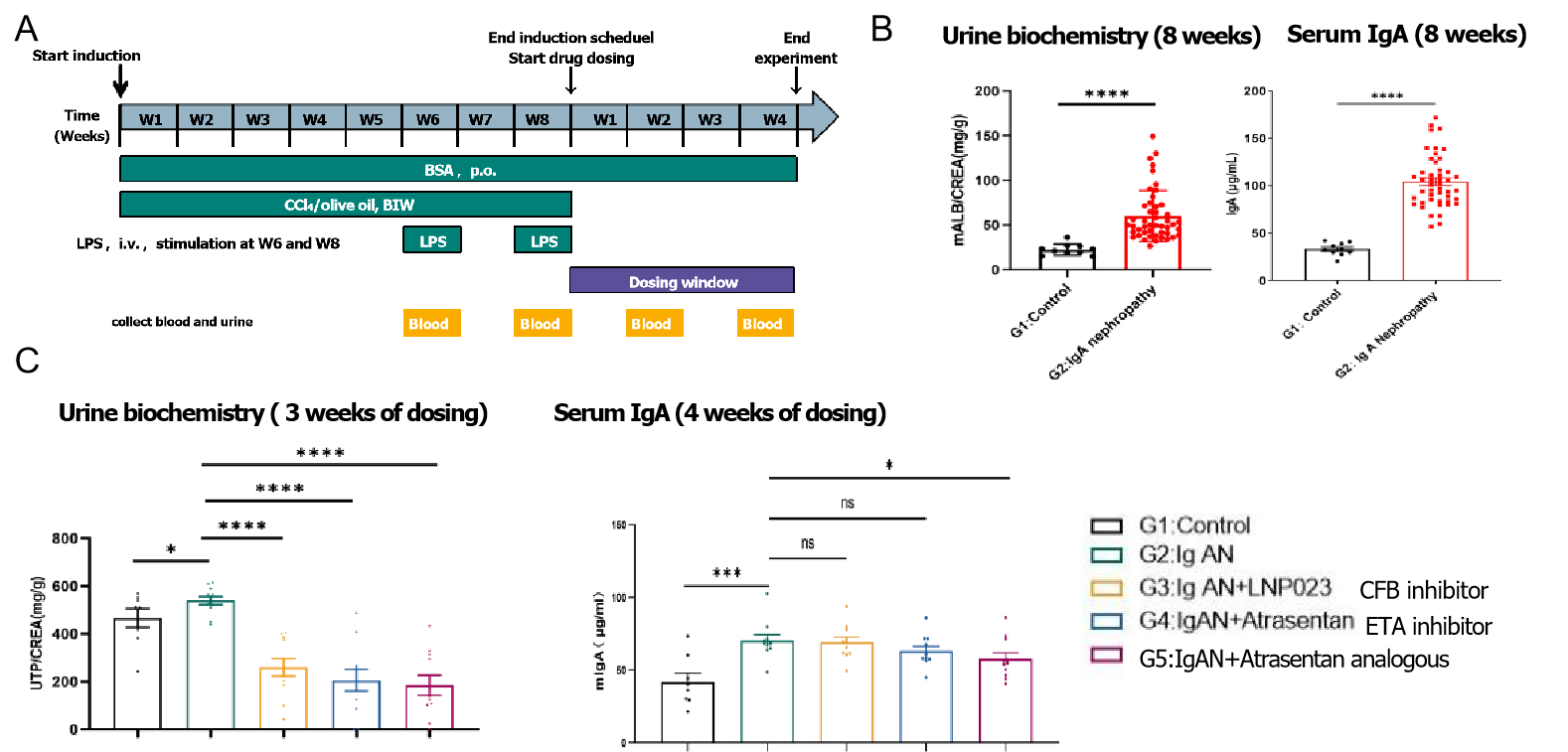
Fig1. The efficacy study of Test Article on BSA+CCL4+LPS induced IgAN mouse model. BALB/c, female, 6-8 weeks were used in this study (n=8). A. Study design. B. UACR (mALB/CREA) and serum IgA were elevated significantly after induction; C. UTP/CREA reduced following treatment with CFB inhibitor and ETA inhibitor.
In BSA+CCL4+LPS induced IgAN mouse model, after 8 weeks induction and 4 weeks treatment, CFB inhibitor and Atrasentan improved the development of disease accompanied with the significant decrease of UTP/CREA.
Note: No pathological change in kidney in this model were observed in accordance with references.
CD89-IgA humanized spontaneous IgAN model
Excessive inflammation caused by IgA immune complex may lead to autoimmune diseases, such as immunoglobulin A(IgA) nephropathy (IgAN). It has been proposed that in IgAN there is specific pIgA-induced shedding from myeloid cells into the circulation, and that released sCD89–pIgA complexes amplify the molecular size of circulating IgA-immune complexes (IgA-ICs) which promote IgA deposition in the mesangial, contributing to IgAN development. We developed CD89-IgA double humanized mice for IgAN study.
Survival rate and proteinuria level

Spontaneous death and proteinuria leakage were observed in female B6-Cd14-hCD89/hIGHA1 mice.
IgA level in serum
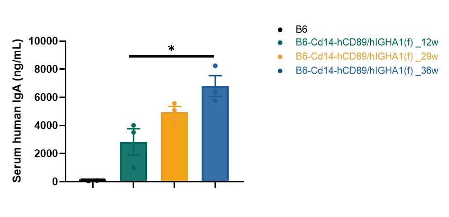
The expression of human IgA1 in the serum of female B6-Cd14-hCD89/hIGHA1 mice can be detected, which continued to increase gradually over time
Pathological analyses

Notes: Increased mesangial cells (green arrow) and deposition of mesangial matrix (blue arrow)
The first row: bar = 100μm; The second row: bar = 20μm
Pathological analyses were performed using H&E and PAS staining. Female B6-Cd14-hCD89/hIGHA1 mice exhibited the phenotypic characteristics of IgAN characterized by the presence of basement membrane thickening, inflammatory cell infiltration, fibrous tissue proliferation, increased mesangial cells and deposition of mesangial matrix.
IgA deposition in kidney
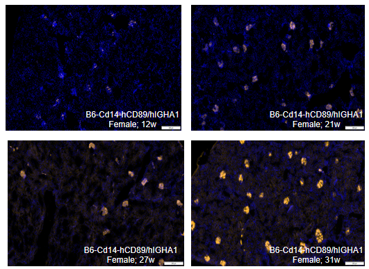
Human IgA deposition in the kidney was detected by immunofluorescence staining using an anti-human IgA antibody in B6-Cd14-hCD89/hIGHA1 mice. With mice increase in age, the amount of human IgA deposition detected increased.