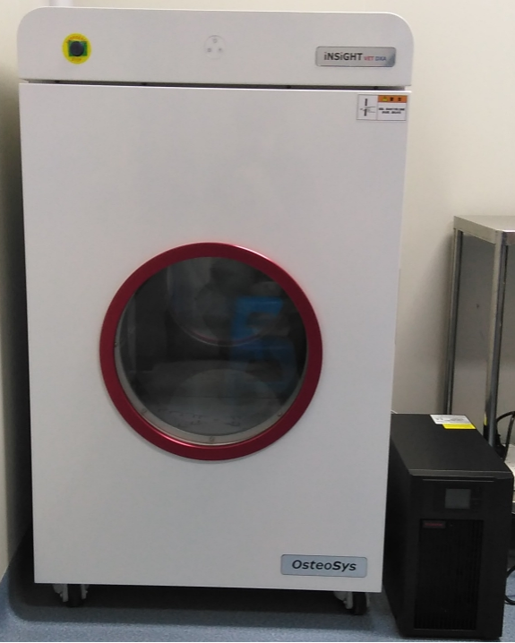
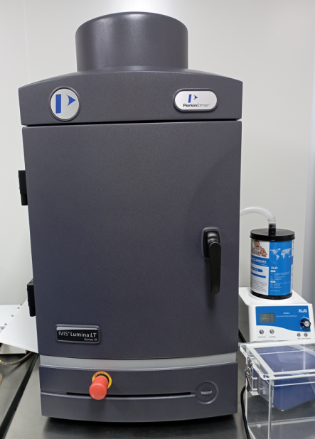
The GemPharmatech Imaging and Analysis Platform is equipped with a dual-energy X-ray absorptiometry and an in vivo optical imaging system. The dual-energy X-ray absorptiometry provides high-resolution X-ray imaging for assessing bone density and body composition in live animals and ex-vivo tissues, primarily accommodating model organisms such as mice and rats. It's used for research on diseases such as osteoporosis, obesity, metabolic disorders, and aging.
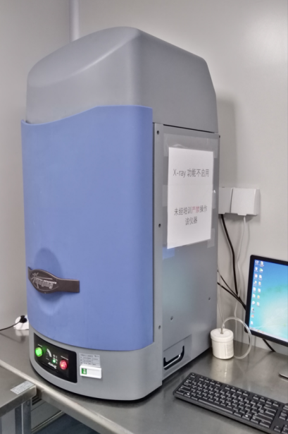
The in vivo optical imaging system at GPT offers high-quality fluorescence and bioluminescence imaging capabilities, enabling tracking and detection of labeled cell and gene activities in vivo. It's also used for studying the distribution, metabolism, targeting, and accumulation of luminescent substances such as fluorescent probes, novel luminescent materials, and labeled drugs in vivo.
Service Offering
(1) Dual-Energy X-ray Absorptiometry
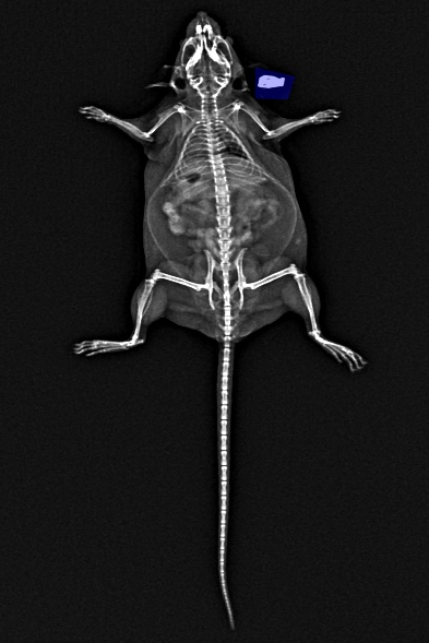
The system utilizes high-energy and low-energy X-rays to calculate body composition by measuring the differential attenuation within the examined body. It provides high-resolution X-ray scan images used to measure whole-body or local bone density, as well as fat and lean tissue content in experimental animals.
(2) In vivo Optical Imaging System
The in vivo optical imaging system collects and analyzes light signals, enabling quantitative monitoring of tumor growth, metastasis, drug distribution, and other physiological signals in mice. This system facilitates drug efficacy evaluation and detection of drug distribution.
GPT Advantages
(1) Dual-Energy X-ray Absorptiometry
Precise bone density detection
Safe and convenient
Reliable data
High image quality
(2) In Vivo Optical Imaging System
Large field of view
High throughput
Multimodal imaging
Absolute quantitative analysis
Case Studies
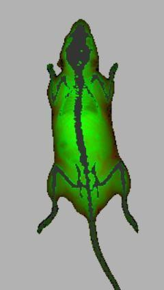
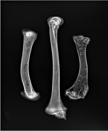
(1) Dual-Energy X-ray Absorptiometry: can export X-ray images, bone mineral density images, and pseudo-colored body composition images
X-ray images (for measuring partial bone length, determining fractures, and locating implants using high-resolution images).
BMD Images

Body composition images (reflecting the distribution and changes in body composition, with red indicating fat distribution)
Isolated bone tissue
(2) In Vivo Optical Imaging System
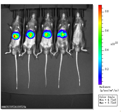
Drug distribution experiment: Four mice were treated with a fluorescence-labeled drug, while the fifth served as a control.
