Asthma is a chronic lung disease affecting people of all ages. It is caused by inflammation and muscle tightening around the airways, which makes it harder to breathe. Symptoms can include dry cough, night cough, difficulte breath, shortness of breath, chest pain or tightness, wheezing.
1. OVA-induced asthma model
Experimental mouse strains: BALB/c, Female
Modeling reagent: OVA
Modeling method: intratracheal instillation
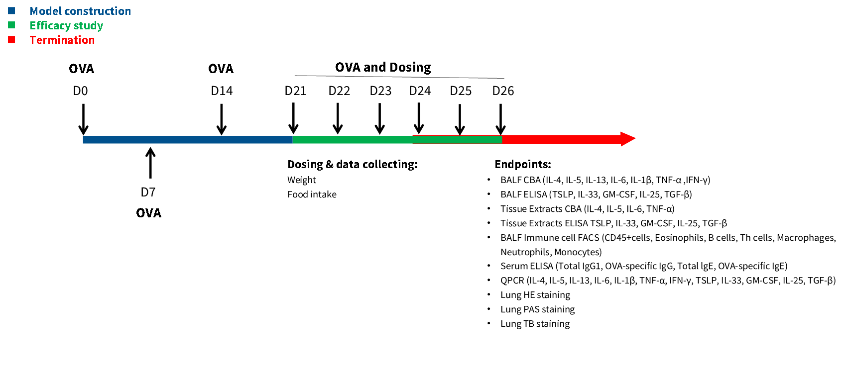
Validation Data
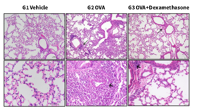
HE Staining. Inflammatory cells were showed by black arrows. Bar=100 μm in the top row. Bar=20 μm in the bottom row.
Compared to G1, G2 showed moderate to massive infiltration of inflammatory cells including eosinophils, mast cells, lymphocytes, macrophages, and neutrophils surrounding the airways and vessels of the lungs. Compared to G2, G3 showed significantly reduced inflammatory cell infiltration surrounding the airways and blood vessels.
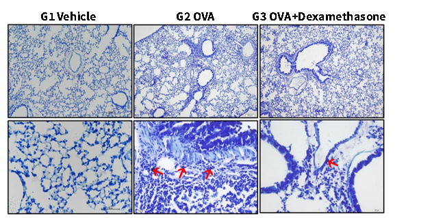
Toluidine Blue Staining. Mast cell degranulation was showed by red arrows. Bar=100 μm in the top row. Bar=20 μm in the bottom row. Compared to G1, G2 showed occasional degranulation of mast cells in the interstitium of the lungs. Compared to G2, G3 showed rare mast cell degranulation in the interstitium of the lungs.
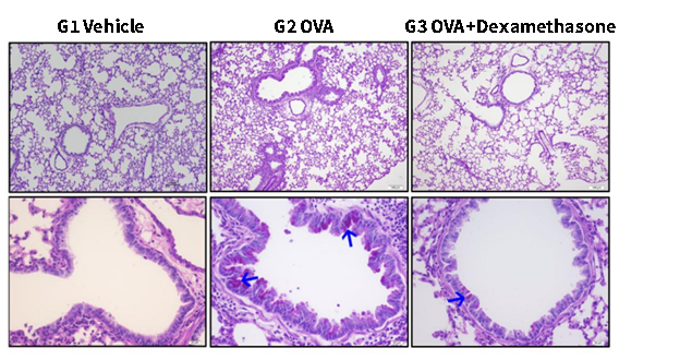
PAS Staining. Mucus attachment was shown by blue arrows. Bar=100 μm in the top row. Bar=20 μm in the bottom row.
Compared to G1, G2 showed moderate to abundant PAS-positive mucous material in the bronchi or bronchiolar mucosal epithelium. Compared to G2, G3 showed only occasional PAS-positive mucous material in the mucosal epithelium of the bronchi or bronchioles.
Serum toal IgE, OVA-sIgG and OVA-sIgE level were decreased in OVA induced asthma mouse model by DXSM(Dexamethasone).
2. OVA-induced asthma model
Experimental mouse strains: BALB/c, Female
Modeling reagent: OVA
Modeling method: Oropharyngeal Intratracheal (OA)
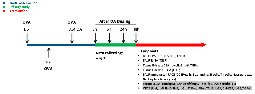
Validation Data

The TSLP level in lung tissue significantly increased after 3 hours of OVA induction.
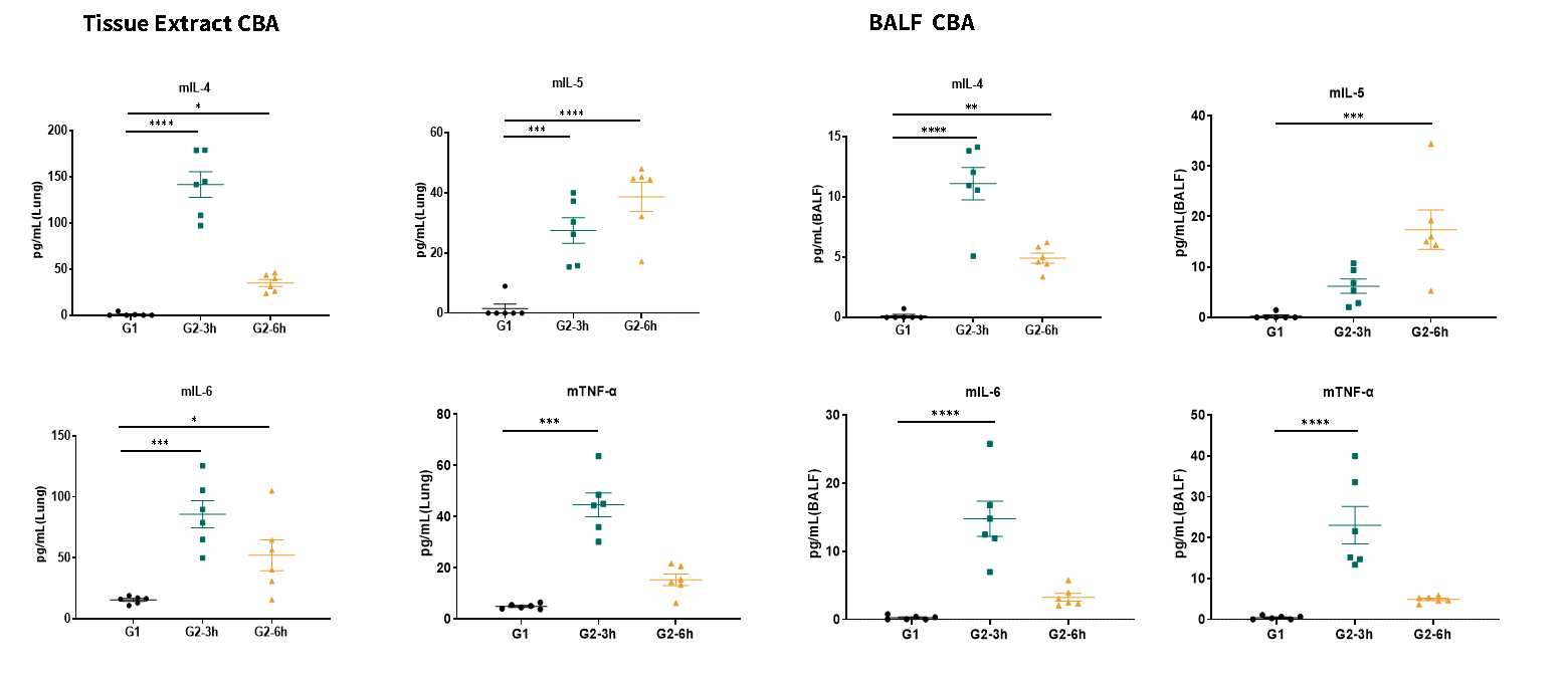
IL-4, IL-5, IL-6 and TNF-α level increased significantly after OVA induction.
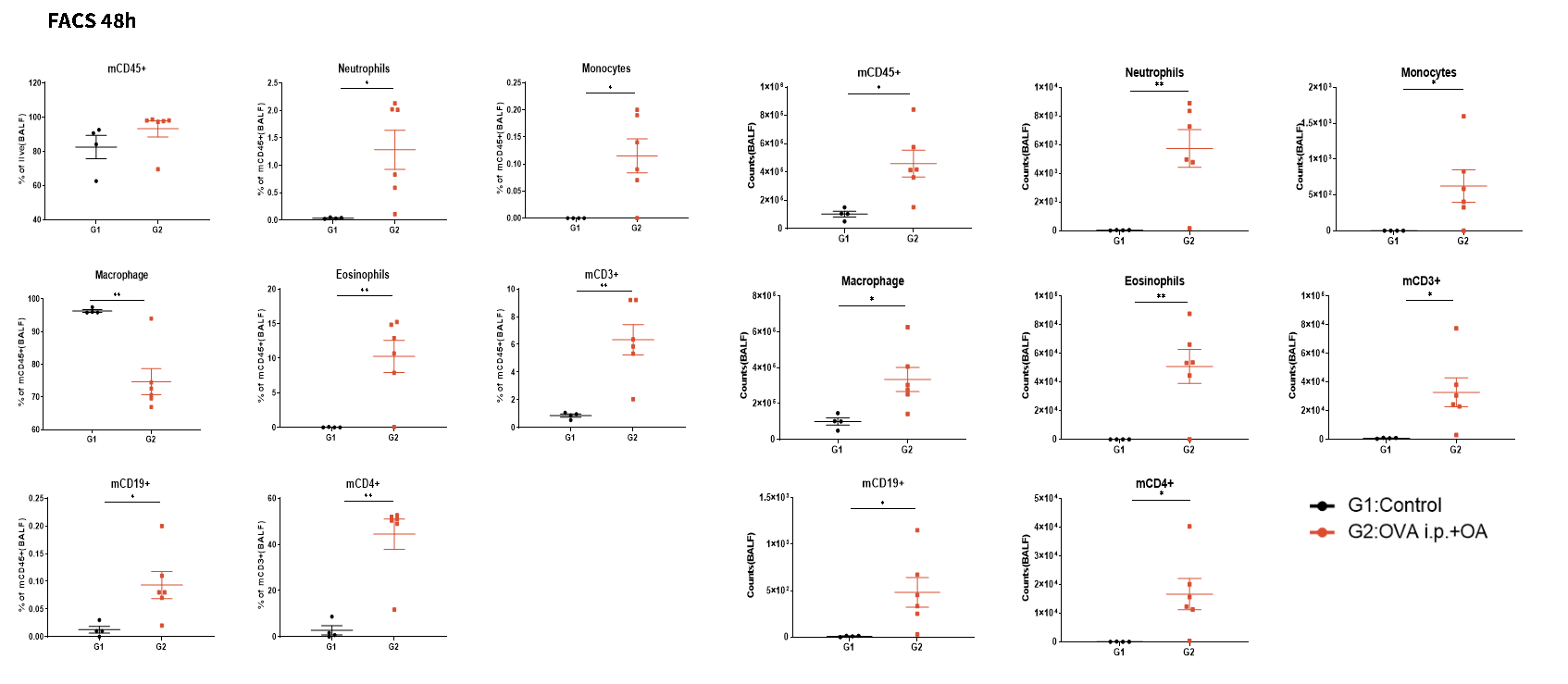
BALF mCD45+ cells, B cells, Th cells, neutrophils, eosinophils, macrophages, monocytes level increased significantly after OVA induction
3. HDM-induced acute asthma model
Experimental mouse strains: BALB/c, Female
Modeling reagent: HDM
Modeling method: intranasally, i.n.
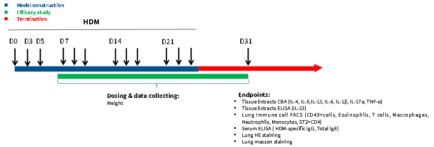
Validation Data
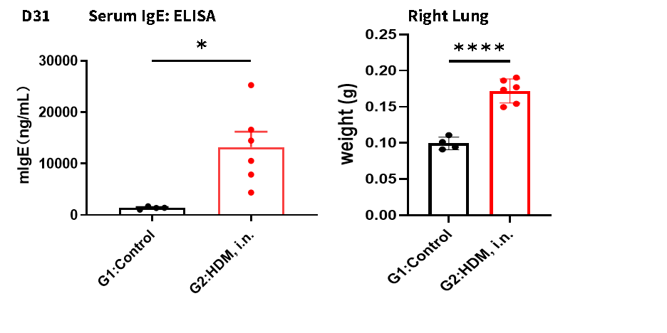
IgE increased significantly after HDM induced, enlarged lung tissue was observed in HDM induced group.

Bronchial epithelial goblet cells (orange arrows), multinucleated giant cells (green arrows). Lung HE score and airway wall thickness increased in HDM induced group.
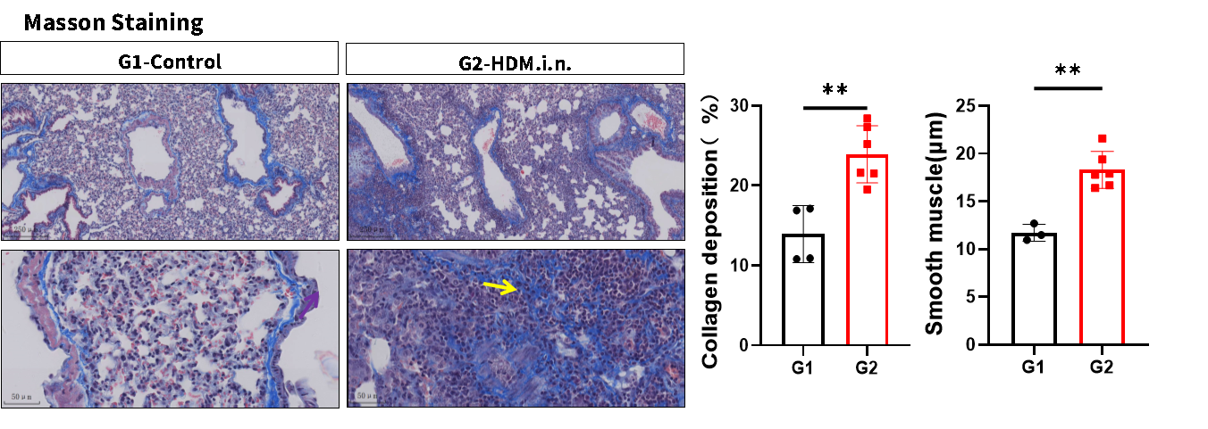
Collagen fibers around alveoli (yellow arrow), Smooth muscle was thickened and collagen fibers proliferated in HDM induced group.


