

Ferric Chloride-induced Carotid Artery Thrombosis
Carotid artery thrombosis refers to the pathological formation of blood clots within the carotid arteries, representing a major cause of ischemic stroke. This condition typically develops through three key mechanisms: rupture of atherosclerotic plaques, endothelial dysfunction, or blood hypercoagulability. The resulting thrombi can either partially or completely obstruct blood flow, leading to cerebral hypoperfusion and neurological deficits.
The ferric chloride (FeCl₃)-induced carotid artery thrombosis model is a widely used preclinical approach for studying arterial thrombosis and evaluating antithrombotic therapies. In this model, a filter paper saturated with FeCl₃ solution is applied topically to the exposed mouse carotid artery, inducing oxidative endothelial injury that triggers rapid thrombus formation. FeCl₃ generates reactive oxygen species, causing endothelial denudation, platelet adhesion/activation, and fibrin deposition, mimicking human atherothrombosis. Real-time blood flow is measured using Laser Speckle Contrast Imaging (LSCI), allowing quantification of occlusion time and thrombus stability.
The FeCl₃-induced carotid artery thrombosis mouse model is a reproducible and efficient preclinical tool for studying arterial thrombosis and evaluating antithrombotic therapies. By generating oxidative endothelial injury, it rapidly initiates platelet activation and thrombus formation, closely mimicking human atherothrombotic events and is suitable for screening of antiplatelet/anticoagulant drugs.
Example Data
Laser Speckle Contrast Imaging and quantification of blood flow
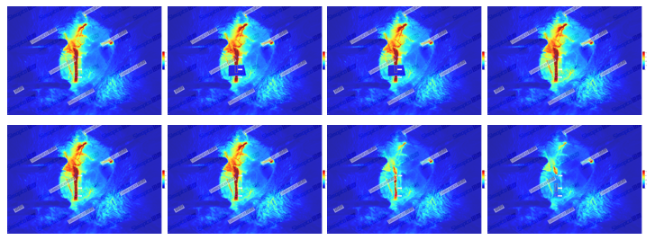
Figure 1. Analysis of carotid blood flow after FeCl₃ filter incubation. LSCI demonstrates progressive vascular occlusion following ferric chloride application: Blood Flow Dynamics (Upper Panel) Initial high perfusion (red/yellow in ROIs). In this assay, Thrombus formation occurs within 5-15 minutes post-application, with complete occlusion typically observed by 30 minutes. Different ferric chloride (FeCl₃) concentrations impact thrombus formation efficiency.
Quantification of blood flow
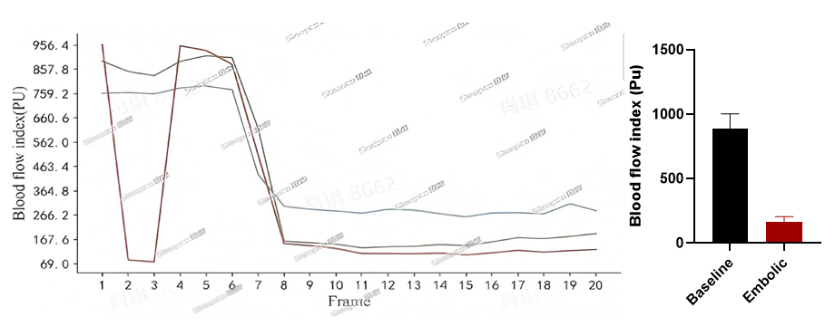
Figure 2. Quantification of LSCI in FeCl₃-Induced carotid thrombosis model. Gradual flow reduction across frames (5 mins/frame). Near-complete flow cessation by Frame 8. Quantitative analysis show Baseline flow: 956.4 PU (Frame 1), background signal: 60 PU. The flow sharply declines to 167.7 PUat Frame, showing 89.3% flow reduction after surgery 20 minutes. This result indicates successful occlusion in FeCl₃-Induced carotid thrombosis model.